GOAL: Relieve anterior scalene spasm
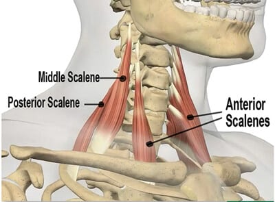
Electricians, painters, dancers, and competitive athletes such as swimmers and pitchers may develop anterior scalene syndrome due to excessive frictioning of cervical nerves as they traverse through fibrotic anterior and middle scalene tendons at the interscalene triangle. Symptoms may present in the anterior neck, chest wall or down into the arms and hands sometimes making it difficult to hold onto objects or to reach overhead. Prolonged anterior and middle scalene spasm due to injury, poor posture or overuse can also cause residual problems as they tug on the first rib pulling it up against a drooping clavicle. When the brachial plexus gets squashed between the clavicle and rib, a condition termed ‘costoclavicular syndrome’ arises. This disorder is one of the leading contributors to thoracic outlet syndrome (TOS).
Because the shoulder and neck area consist of very complex body parts with muscles and connective tissues going in all possible directions, therapists must focus intent on correctly assessing postural strain patterns prior to ‘chasing the pain’. A few commonly applied provocation tests for assessing TOS such as the Adson maneuver (scalenes), ‘Hands-up’ (pec minor), Allen (radial pulse) and the Elevation maneuver for costoclavicular canal impingement may be used. These tests may or may not momentarily reproduce symptoms, but are sometimes helpful in ruling out other causes which may produce similar symptoms.
Due to the overlapping of symptoms, it’s often difficult to make a definitive assessment using provocation tests. Fortunately, advancements in nerve imagery using Magnetic Resonance Neurography are providing more accurate monitoring of exact sites of peripheral nerve damage. These tests, accompanied by thorough history and posturofunctional evaluations, are extremely helpful for massage and functional movement therapists when it comes to treating entrapment sites that irritate the nerve bundle and bring sensation to the arm and hand.
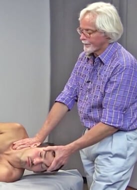
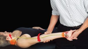
Therapeutic outcomes reap greater rewards when the nerves along the entire length of the arm are examined and treated beginning proximally from the cervicothoracic spine down through the wrist and hand. In the accompanying video, I demonstrate a very effective anterior scalene maneuver for relieving brachial plexus compression. Those of you who have attended my workshops or have taken my Technique Tour home-study course have seen how beautifully this maneuver fits into our neural mobilization routines from the Treating Trapped Nerves course.
When learning anterior neck work, special caution must be taken to avoid placing finger pressure on all neurovascular structures. Therefore, I suggest you practice the following in a supervised setting.
Anterior Scalene Technique (Image 2.)
GOAL: Release adhesions in anterior scalene muscles
ACTION: (Client left sidelying)
- Therapist helps client left rotate their head to expose clavicular (lateral) border of SCM
- Therapist’s left hand controls the client’s head and his right extended soft finger pads gently slide under SCM
- Therapist’s left hand slowly rotates client’s head and neck back to neutral and in to hyper-extension while fingers of right hand “scrubs” the anterior tubercles (transverse processes)
- Therapist repeats the entire maneuver as gently curled fingers inspect for scalene fibrosis along anterior tubercles (C2 to C6)
- If thickness or knots are palpated, client deeply inhales or tucks (and releases) chin as therapist holds sustained gentle pressure until GTO receptor release is felt
- Repeat 3 to 5 times and retest neck flexion firing order
On sale this week only!
Save 25% off the Upper Body Course!
NEW! Enhanced video USB format!!!
Learn unique approaches to work with the shoulder girdle, arms, neck, and torso, this course will prepare you to relieve painful myoskeletal issues in the upper body. Through video demos and animation, you’ll learn to identify several compensatory movement patterns and their associated reflexogenic pain. With this understanding of where the true source of problems arise, you’ll be able to develop highly effective treatment protocols and deliver lasting results. This course provides you with the skills you need to confidently relieve pain issues in the upper body. (20 CE)
Bonus: When you order the Home Study version, get the eLearning course and and an additional 2CE Ethics eCourse for free!
Save 25% off the Upper Body Course this week only. Offer expires Sunday, March 1st. Click the button below for more information and to purchase the course for CE hours and a certificate of completion to display in your office.

